Ankle and Foot Anatomy Basics
Basic anatomy for any joint structure within the body includes bones, joints, muscles, tendons, and ligaments. For our purposes, we will be discussing anatomy of the ankle and foot structures specifically.
Terms to Know:
- Lateral: outside
- Posterior: backside
- Anterior: frontside
- Medial: inside
- Distal: situated away from the center of the body
- Proximal: situated closer to the center of the body than distal
Anatomy of a Joint Structure
A joint is a part of a body where two or more bones meet. The ends of these bones are covered by Cartilage. To define, Cartilage is a connective tissue structure that helps provide shock absorbing properties when performing activities. In addition to cartilage, Synovial Fluid presents within each joint space and promotes smooth movement of the joint. There are also important connective tissues called Tendons and Ligaments that make up each body structure. A tendon is a tissue that connects muscle to bone. Similarly, ligaments connect bone to bone.
Ankle Anatomy
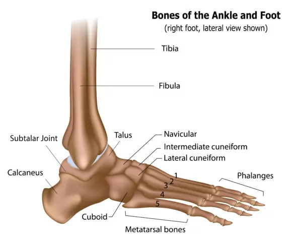
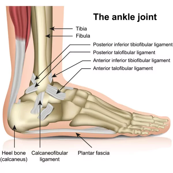
The ankle joint is formed where the bones of the lower leg, the Tibia and Fibula, meet the Talus. With this, the portion of the fibula, located at the ankle level, is referred to as the Lateral Malleolus while the portion of the tibia at the ankle level is referred to as the Medial Malleolus. These two (2) ankle bones are commonly fractured in injuries. Additionally, just below the ankle joint is the Subtalar Joint, which is located between the Talus and the Calcaneus. The Calcaneus is also known as the heel bone.
Each of these joint junctions are responsible for allowing movement of the ankle in four different directions: Plantarflexion, Dorsiflexion, Inversion, Eversion. To demonstrate these directions, point your foot. Your ankle is in the Plantarflexed position. Now, bring your toes towards your head. This is the Dorsiflexion position. From here, if you move your toes inwards you will be in the Inverted position. Alternatively, if you move your toes outwards, you will be in the Everted position.
In many cases, normal wear and tear from aging can predispose the ankle joint to arthritis. However, wearing appropriate footwear for your activities and performing ankle strengthening and stretching exercises can delay the onset of arthritis. Tune into our blog Osteoarthritis vs Rheumatoid Arthritis to learn more about the causes of arthritis and ways to prevent or delay its onset.
Muscular and Tendon Anatomy of the Ankle
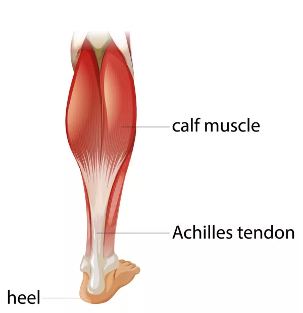
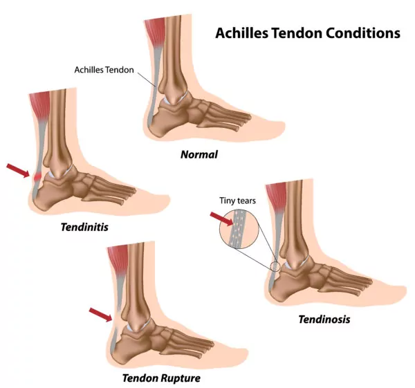
The posterior side of the lower leg houses the calf muscles. These muscles attach to the Achilles tendon, which is the largest tendon in the body. This is exposed to large amounts of force in activities such as running or jumping, making it prone to injury. The Tibialis Posterior muscle also lives in the posterior side of the lower leg. Continuing, the tendon associated with this muscle crosses over the middle portion of the ankle and is called the Posterior Tibialis Tendon. Those with flat feet may be at risk for additional strain on this tendon, which if not addressed, can lead to tendonitis.
The lateral compartment of the lower leg contains two muscles, the Peroneal Brevis and the Peroneal Longus muscles. Subsequently, the tendons of these muscles travel on the outside of the ankle and can also be subject to strain with overuse. Other important structures over the lateral ankle include three (3) lateral ligaments: the Anterior Talofibular Ligament (ATFL), the Calcaneofibular Ligament (CFL), and the Posterior Talofibular Ligaments (PTFL). These structures are vital for stability of the ankle. Injury to these ligaments, as commonly seen with ankle sprains, can lead to long-term instability if not treated properly. Visit our blog What to do for a Sprained Ankle to learn more about how to treat an ankle sprain.
Foot Anatomy
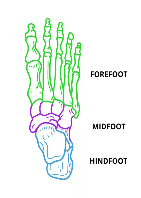
The feet each contain 26 bones as well as many soft tissue structures including tendons, ligaments, nerves, and vascular structures. In medical discussion, the foot is often broken down into three (3) portions: the Hindfoot, Midfoot, and Forefoot.
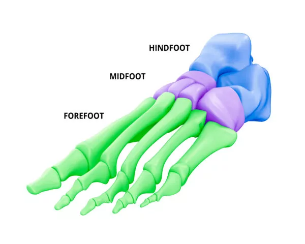
The Hindfoot
The Hindfoot contains the talus and calcaneus, otherwise known as the ankle bone (Talus) and heel bone (Calcaneus). As mentioned previously, these two (2) bones join to make up the Subtalar Joint and allow the foot to rotate in multiple directions at the ankle level. Additionally, the Plantar Fascia Tendon runs from the calcaneus to the end of the metatarsal bones located in the forefoot.
The Midfoot
The midfoot houses the five (5) tarsal bones and three (3) ligaments. These ligaments are referred to as the Lisfranc Joint Complex and connect the midfoot to the forefoot. The five metatarsal bones, to include the navicular, cuboid, and three (3) cuneiform bones, all help to form the arches of the foot. Alone, the midfoot bones do not provide all the stability for the midfoot. The Midfoot Ligament Complex is also responsible for providing stability. This complex of Lisfranc ligaments includes the Dorsal Lisfranc Ligament, the Interosseous Lisfranc Ligament, and Plantar Lisfranc Ligament. These ligaments help maintain alignment of the tarsal and metatarsal bones and act as shock absorbers during activity.
The Forefoot
Within the forefoot, you will find the metatarsals and the toe bones, medically known as the Proximal, Medial, and Distal Phalanges. The Distal Phalanges are located at the end of each toe while the Proximal Phalanges are situated closest to the metatarsals and allow the toes to bend. You will also find the four (4) deep transverse metatarsal ligaments within the forefoot. These ligaments help to stabilize the metatarsal bones and prevent the foot’s arch from widening or collapsing. These forefoot structures are key to navigating various surfaces and maintaining balance when performing activities such as walking, running, pivoting, or jumping.
Due to their weightbearing capacities and vital function in performing day-to-day activities, the feet and ankles can be subject to many different conditions that may cause discomfort. Because of this, Arthritis in the joints of the feet can occur secondary to routine wear and tear or following injuries. Additionally, different deformities such as flat feet, high arches, bunions, hammertoes, or mallet toes may occur. These deformities can result in pain, calluses, and difficulty wearing shoes. If you are experiencing any of these symptoms or conditions, please give our office a call and we will design a treatment plan based on your personal needs and get you back to the lifestyle you enjoy.
The intrinsic anatomy of the ankle and foot can cause various injuries or conditions to become very complex. For more information on conditions of the foot and ankle and treatment options available, check out our blog series developed by our providers within the Foot and Ankle Center.
Developed by the Colorado Springs Orthopaedic Group Foot and Ankle Center
Meet Our Providers


G. Alex Simpson, DO