Anatomy of the Knee
A comprehensive guide detailing the bones, muscles, tendons, and ligaments of the knee
Running, jumping, walking, hiking, our knees support us through it all. To help provide these high-impact functionalities, the knee’s structure is one of the most complex and critical joints within the human body. Let’s take a closer look at the anatomy of the knee to better understand the knee and its many functionalities.
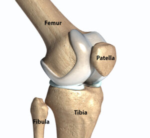
Bones of the Knee
The knee joint is formed by the femur (thighbone), the tibia (shinbone), and the patella (kneecap). The femur is the largest bone in the body and it forms the upper part of the knee. The tibia forms the lower part of the knee and is the weight-bearing bone of the lower leg. The patella is a small, flat bone located in front of the knee. It slides along a groove in the femur as the knee bends and straightens.
Ligaments of the Knee
Within the knee, we have four primary ligaments: the ACL, PCL, LCL, and MCL. Ligaments are strong, fibrous tissues that connect bo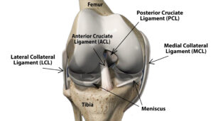 ne to bone and provide stability within the joint.
- The Anterior cruciate ligament (ACL) is located in the center of the knee. The ACL helps prevent the tibia from moving forward excessively.
- The Posterior cruciate ligament (PCL) is located in the center of the knee. The PCL helps prevent the tibia from excessively moving backwards.
- The Lateral collateral ligament (LCL) is located on the outside of the knee. The LCL helps prevent the knee from bending outward too an excessive degree.
- The Medial collateral ligament (MCL) is located on the inner most side of the knee. The MCL helps to prevent the knee from bending inward to an excessive degree.
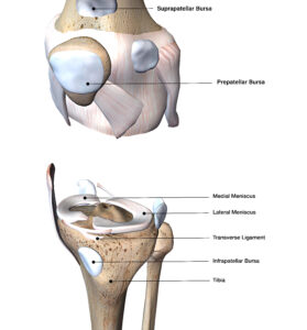
Bursae of the Knee
Bursae are small, fluid-filled sacs. They can be found in many areas of the body and serve as friction reducers for the tissues surrounding a joint. In the knee, we have three primary bursas: The Suprapatellar bursa, the Prepatellar bursa, and the Infrapatellar bursa.
Menisci of the Knee
Between the Femur and Tibia, there are two cushion like structures. These structures are referred to as the Menisci, or Meniscus in singular context. The Menisci act as shock absorbers when performing activities such as running, jumping, walking, etc. and help to distribute our body weight evenly across the knee joint.
Tendons of the Knee
Let’s start by addressing what tendons are and what they do. Tendons are fibrous tissues that connect muscles to bones and help initiate movement as our muscles contract and relax.
For example, as you take a step forward, your quadricep muscles contract. The quadriceps tendon that’s attached to the quadricep muscles then helps initiate movement within the patella, the patellar tendon and tibia so that the entire lower leg moves forward in congruency.
You may have heard of a condition called Patellar Tendonitis. This refers to an injury of one of the primary tendons within the knee, the Patellar Tendon. The Patellar Tendon connects the patella to the tibia and with the help of the quadriceps tendon, is responsible for extending the knee when you kick, run or jump. It is one of the most injured tendons especially amongst those who frequently run or jump on hard surfaces.
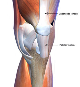
The second most primary tendon within the knee is the Quadriceps Tendon. This tendon connects the quadricep muscles to the patella and in conjunction with the patellar tendon, helps to extend or straighten the knee.
To recap Tendons vs Ligaments:
- What they connect:
- Tendons connect muscles to bones
- Ligaments connect bone to bone
- What they do:
- Tendons help initiate movement between muscles and bones
- Ligaments help hold structures together and provide stability within those structures
The Iliotibial Band (The IT Band)
You’ve probably heard of the IT band before. But what is it and what does it do? The IT band is a band of thick fibrous tissue that runs from the hip down to the lateral aspect of the Tibia. This band of tissue helps to provide stability to both the knee and hip and as it runs down to the top of the Tibia, helps to prevent dislocation of the knee as well.
Muscles of the Knee
The knee is surrounded by several muscle groups, each of which help maintain stability and move the knee joint through motion in conjunction with ankle and hip joint movements when performing daily activities.
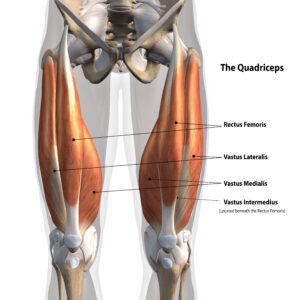
The Quadriceps
Starting from the top and front (anterior) side of the knee, we have the four quadricep muscles that attach at the base of the femur, just above the knee joint:
- The Rectus Femoris
- The Vastus Lateralis
- The Vastus Medialis
- The Vastus Intermedius
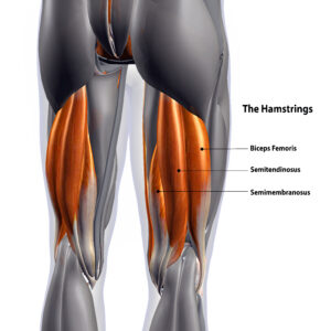
The Hamstrings
On the back (posterior) side of the leg are the three hamstring muscles:
- The Biceps Femoris
- The Semimembranosus
- The Semitendinosus
The hamstrings attach at the base of the femur and top of the tibia. If you touch the back of your knee, you may be able to feel where these insertion points are located. The hamstrings are the muscles that control flexion or bending of the knee and help to stabilize the knee when it’s extended (when the quads are contracted and the leg is straightened). They also assist in turning the knee inwards (referred to as internal rotation) and outwards (referred to as external rotation).
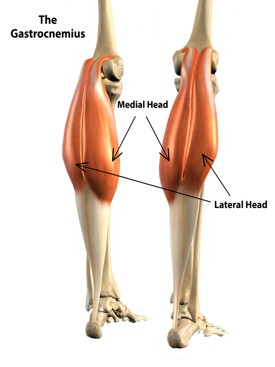
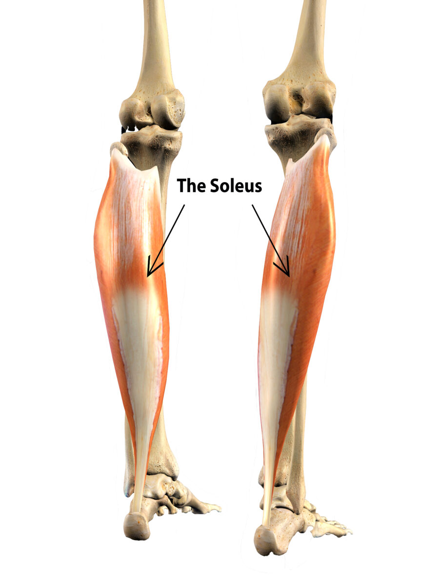
The Calf Muscles
Moving down to the lower part of the knee, we have the Anterior Tibialis on the front side and the Gastrocnemius and Soleus muscles on the back side.
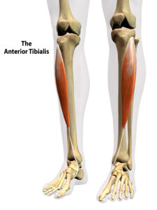
The Anterior Tibialis originates at the top of the tibia and is responsible for deceleration during activities. When strengthened appropriately, it reduces the amount of force our knees experience when slowing ourselves down. An easy way to remember the Anterior Tibialis is to remember ‘anterior’ refers to the front side. So the Anterior Tibialis is on the front aspect of the tibia (shin bone).
Moving to the back or posterior side of the lower leg, the Gastrocnemius has two muscle heads, the medial (inner) head, and the lateral (outer) head. Both of which originate on the back of the femur and run down to the Achilles tendon. Similarly, the soleus muscle originates at the base of the femur, runs down to the heel bone via the Achilles tendon, and lies underneath the lateral head of the gastrocnemius. Both the Soleus and Gastrocnemius (gastroc for short) support the hamstrings in knee flexion and provide stability to the knee when you jump, run, flex or extend the knee during activities.
The anatomy of the knee is a very complex but with these complexities come numerous functionalities. These features make the knee truly one of the most amazing joints and allow us to perform so many daily activities from walking up stairs, to hiking, biking, and everything between. It is however, important to note that with great complexities, comes great need to take care of these joints as the risk for injury to any of the structures within is increased substantially.
Tune into our knee blogs to learn more about the knees, some of the most common injuries and conditions we see here at Colorado Springs Orthopaedic Group, injury prevention techniques, and when it’s recommended to see an orthopaedic specialist for further evaluation.
If you or a loved one are struggling with chronic knee pain, give us a call to schedule an initial evaluation. Our board certified and fellowship trained orthopedic specialists want nothing more than to see you back doing the things you love to do.
Visit our providers page to meet our specialists.